Executive Summary: The Evolving Landscape of Extremity Deformity Correction
Extremity deformity correction represents one of the most sophisticated, biomechanically demanding disciplines within modern orthopedic surgery. Addressing complex congenital, post-traumatic, metabolic, and developmental skeletal deformities—such as blount's disease, genu varum/valgum, limb length discrepancy (LLD), bone loss from severe open fractures, and non-unions—requires an extraordinary synergy between biological repair mechanisms and mechanical stability.
At the center of successful surgical outcomes is Extremity Deformity Correction Hardware. Modern deformity correction systems have evolved beyond basic Ilizarov pin-and-ring constructs into hyper-precise, software-guided circular spatial frames (hexapods), low-profile anatomical locking plates, and magnetically actuated motorized intramedullary lengthening nails. Global procurement managers and orthopedic hospital purchasing officers face complex decisions when sourcing hardware: balancing biomechanical rigidity, patient comfort, pin-tract infection mitigation, software interface usability, radiolucency, and strict compliance with international regulatory frameworks such as EU MDR 2017/745 and US FDA 510(k).
When selecting extremity deformity correction hardware, evaluating static load capacity alone is insufficient. Procurement decisions must consider the system's dynamic stiffness under physiological weight-bearing, software web-accessibility, multi-planar correction accuracy (Center of Rotation of Angulation - CORA), and compatibility with osteoinductive biological matrix therapies (such as Accell Bone Matrix) to accelerate distraction osteogenesis.
Biomechanical Principles of Distraction Osteogenesis & Deformity Correction
Understanding the clinical efficacy of extremity deformity correction hardware requires a comprehensive grasp of distraction osteogenesis—the physiological process where living bone is incrementally pulled apart (distracted) after an osteotomy to stimulate new bone formation (callus) in the gap. Developed by Dr. Gavriil Ilizarov and refined over four decades by leaders like Orthofix, distraction osteogenesis relies on strict mechanical parameters:
- Latency Period: A post-operative resting phase of 5 to 10 days before distraction begins, allowing initial inflammatory callus formation.
- Distraction Rate & Rhythm: Standard distraction occurs at 1.0 mm per day, typically divided into four 0.25 mm increments per day. Hardware must permit precise micro-adjustments without play or mechanical hysteresis.
- Consolidation Phase: The period where the newly formed woven bone mineralizes into cortical bone. Hardware must maintain total structural stability while permitting micro-motion axial loading (dynamization) to encourage cortical remodeling.
The biomechanical success of any deformity correction assembly hinges on resolving 3D spatial deformities across six degrees of freedom (6-DOF): translation (anterior-posterior, medial-lateral), angulation (flexion-extension, varus-valgus), axial rotation (internal-external), and axial length (distraction-compression). Hardware components must withstand severe torsional, bending, and compressive loads during patient weight-bearing without catastrophic pin-shear or strut deformation.
Comprehensive Hardware Portfolio: Systems Analysis & Recommendations
Orthofix’s global portfolio of extremity deformity correction hardware provides surgeons and hospital procurement networks with modular, multi-faceted solutions tailored to adult and pediatric patient populations.
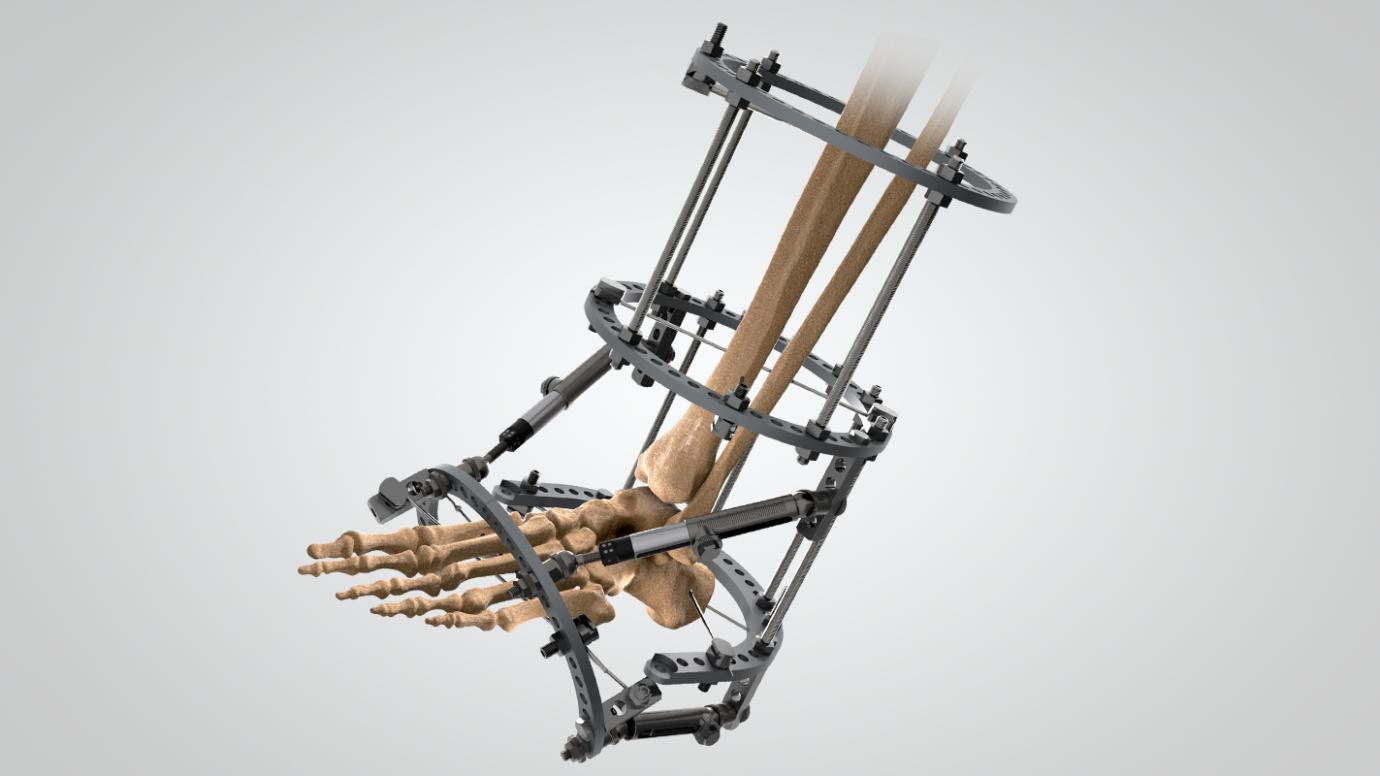
Circular Hexapod Spatial Frames
The gold standard for multi-planar complex deformity correction. Utilizing six telescopic, color-coded struts connecting aluminum-alloy or carbon-fiber rings, hexapod systems solve multi-axis translational, angulated, and rotational deformities via proprietary 3D spatial web software.
- High-grade titanium alloy Schanz screws & HA-coated pins
- Radiolucent carbon fiber full/half rings for unhindered fluoroscopy
- Micrometric strut adjusters for patient-friendly compliance
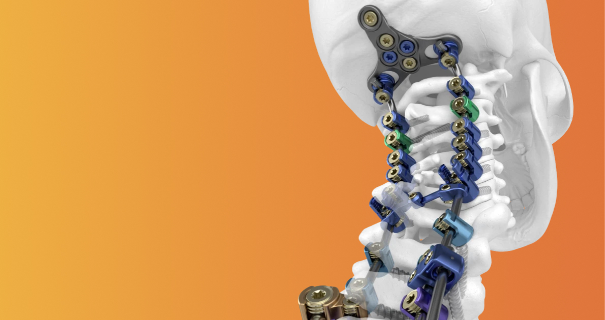
Monolateral Deformity & Rail Fixators
Ideal for femoral and tibial lengthening, post-traumatic bone transport, and single-plane angular correction. Monolateral systems offer a lower profile than circular frames, dramatically improving patient mobility and psychological comfort during prolonged distraction phases.
- Heavy-duty sliding rail clamps with micro-distraction drives
- Self-drilling, self-tapping tapered Schanz screws
- Integrated dynamic spring modules for progressive weight-bearing
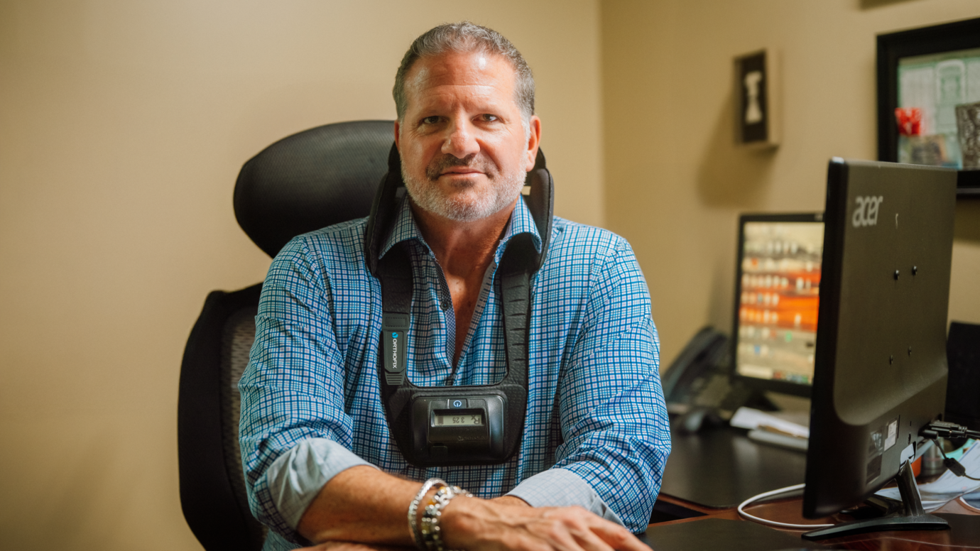
Guided Growth & Anatomical Locking Plates
For pediatric tethering and adult acute deformity correction. Flexible 2-hole tension band plates allow temporary hemiepiphysiodesis to correct coronal angular deformities (genu valgum/varum) in skeletally immature patients without disrupting growth plate viability.
- Low-profile titanium constructs to minimize soft-tissue irritation
- Hinge-screw mechanisms allowing dynamic physeal expansion
- Anatomically contoured periarticular plate options
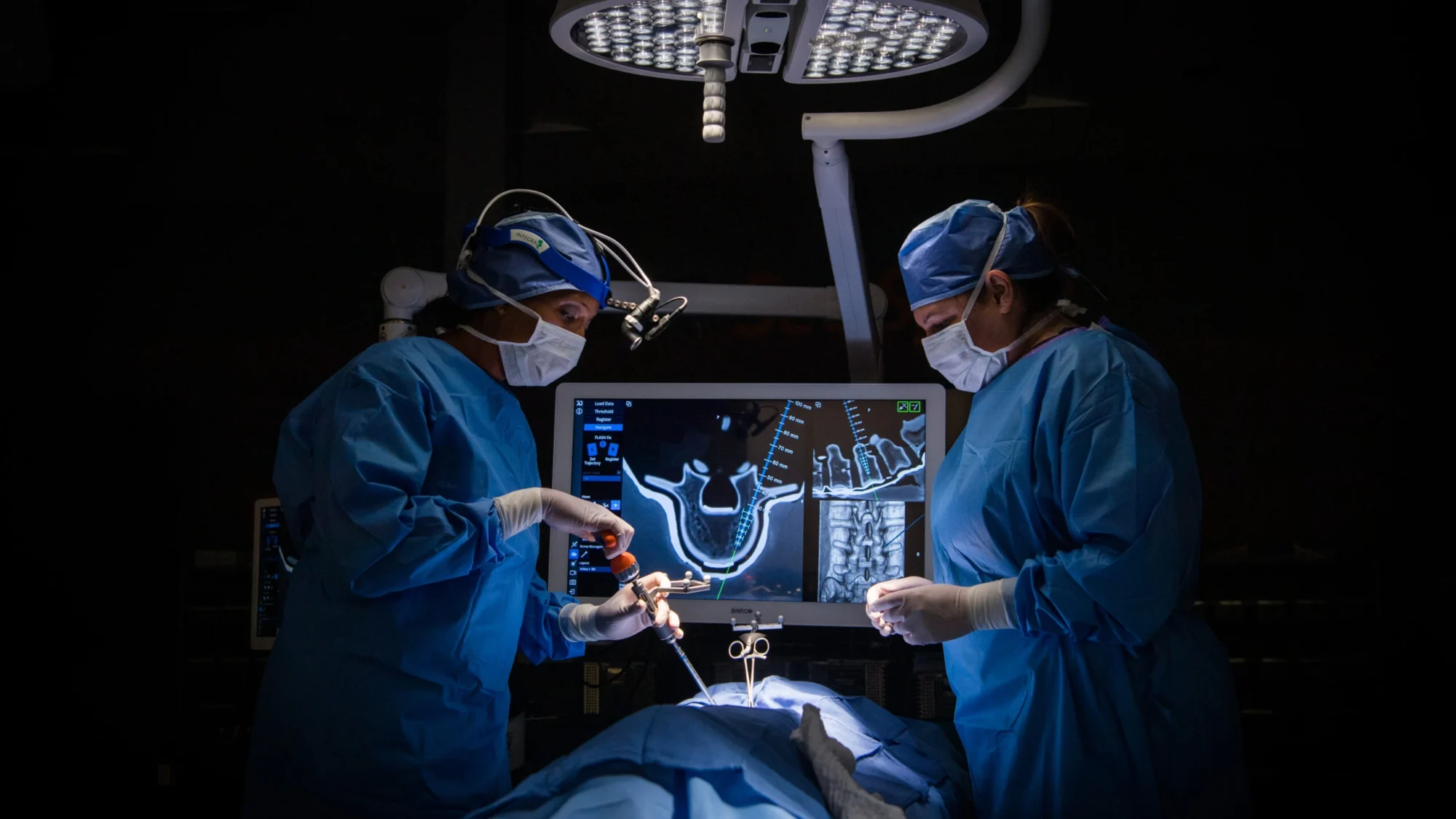
Internal Lengthening & Correction Nails
Next-generation internal limb reconstruction eliminates external pin-tract complications entirely. Utilizing an internal rotor activated by an external hand-held magnetic controller (ERC), these motorized nails allow precise, painless bone lengthening inside the medullary canal.
- Zero pin-tract infection risk and reduced physical therapy barriers
- Hermetically sealed magnetic drive mechanism
- Non-invasive external controller with digital stroke feedback
Technical Specification & Matrix Comparison for Procurement Teams
Global procurement teams must evaluate hardware choices based on rigorous technical criteria, material composition, clinical adaptability, and patient compliance characteristics. The matrix below outlines key parameters across major hardware categories:
| Hardware Architecture | Primary Clinical Indication | Material Composition | Regulatory & Testing Standard | Biomechanical Profile |
|---|---|---|---|---|
| Circular Spatial Frames (Hexapods) | Complex multi-axial deformities, non-unions, severe LLD | Carbon-Fiber / Ti-6Al-4V / Anodized Aluminum | ISO 14602, ASTM F1264, FDA Class IIb | Ultra-high torsional rigidity, 6-DOF adjustment capability |
| Monolateral Rail Systems | Uniaxial femoral/tibial lengthening, bone transport | Aircraft Titanium / Stainless Steel 316L | ASTM F543, ISO 5832-3, CE Mark Class IIb | High bending resistance, low soft-tissue interference profile |
| Guided Growth Plates | Pediatric angular deformity (Genu Varum / Valgum) | Pure Titanium (Grade 4) / Ti-6Al-4V ELI | ASTM F136, ISO 5832-11, FDA 510(k) | Dynamic hinge tensioning, physeal preservation interface |
| Internal Magnetic Nails | Isolated limb length discrepancy, femoral/tibial LLD | High-strength Titanium Alloy (Ti-6Al-4V) | FDA 510(k) Class III, EU MDR 2017/745 | Internal axial loading capacity up to 150 kg; non-invasive distraction |
| HA-Coated Schanz Pins | Percutaneous fixation across all external constructs | Stainless Steel with Hydroxyapatite Coating | ISO 13779, ASTM F1185 | Enhanced pin-bone torque retention, 70% lower infection rate |
Future Procurement Trends & Technological Horizons (2026–2035)
As health systems transition toward value-based healthcare, orthopedic procurement strategies are undergoing a fundamental transformation. Procurement directors are no longer purchasing isolated metal components; they are investing in integrated digital surgical ecosystems. Below are four key trends shaping the future of extremity deformity correction hardware:
1. AI-Driven Pre-Operative Software & Automated Web Planning
Traditional deformity correction required laborious manual radiographic calculations (determining the CORA, landmark angles, and apex of deformity). Modern hardware systems are now paired with cloud-based artificial intelligence algorithms that analyze CT scans and long-leg standing radiographs automatically. These AI engines output exact 3D spatial strut adjustment schedules, reducing surgical planning time from hours to minutes while minimizing human error in strut prescription entry.
2. Patient-Specific 3D-Printed Cutting Guides & Implants
Additive manufacturing (3D printing) using Selective Laser Melting (SLM) in titanium alloy is revolutionary for acute deformity reconstruction. Custom 3D-printed patient-specific osteotomy guides fit precisely onto irregular bony anatomy, allowing surgeons to execute complex multi-planar wedge cuts with sub-millimeter precision before attaching deformity correction hardware.
3. Radiolucent Carbon-Fiber Composites & PEEK Structural Struts
Standard metallic rings and struts cause severe X-ray artifacts, obstructing fluoroscopic visualization of the distraction gap during postoperative follow-ups. Future procurement cycles favor carbon-fiber radiolucent rings and Polyetheretherketone (PEEK) structural struts, enabling crystal-clear radiographic monitoring of bone callus mineralization during the consolidation phase.
4. Tele-Rehabilitation & Smart Digital Telemetry Pin Sensors
Remote patient monitoring is rapidly integrating into extremity deformity correction hardware. Next-generation external struts feature embedded micro-load cells and Bluetooth Low Energy (BLE) transmitters that record real-time axial weight-bearing compliance and pin-site strain. This telemetry alerts surgical teams immediately if a patient over-loads the hardware or misses strut adjustments, drastically lowering failure rates and non-union complications.
Biological Synergies: Accelerating Distraction Callus Formation
Mechanical hardware provides the necessary stabilization, but biological vitality governs bone healing. Orthofix's corporate heritage uniquely bridges hardware engineering with advanced orthobiologics.

Accell Bone Matrix & PEMF Integration
During aggressive limb lengthening or large segment bone transport (e.g., following tumor resection or severe blast trauma), distracted bone gaps risk delayed union or soft-tissue collapse. Combining structural hardware with Accell® Bone Matrix (demineralized bone matrix with reverse phase medium) provides an osteoinductive graft scaffold that accelerates bridging callus formation.
Furthermore, adjunct non-invasive therapies like Pulsed Electromagnetic Field (PEMF) stimulation—found in Orthofix's clinical therapy portfolio—induce cellular upregulation of BMP-2 and BMP-7, shortening consolidation times by up to 35% and allowing faster hardware removal.
Enterprise Advantage & E-E-A-T Credibility: Why Global Hospitals Choose Orthofix
With over four decades of dedicated medical device manufacturing, Orthofix stands as a global pillar of trust, empirical evidence, and clinical rigor. Global procurement partners benefit from unparalleled operational strengths:
44+ Years of Legacy
Founded in 1980 in Verona, Italy, Orthofix pioneered the dynamic external fixator. Our decades of engineering refinement guarantee unmatched biomechanical reliability.
Global Supply Footprint
Active in 70+ countries with localized distribution centers, guaranteeing rapid supply chain response times, stock availability, and instrument kit delivery.
500+ Clinical Studies
Our extremity hardware is backed by extensive peer-reviewed literature, clinical trials, and biomechanical validation in top-tier orthopedic journals.
World-Class Medical Education
We host global surgical workshops, cadaveric labs, and hexapod software training courses to ensure operating room excellence and patient safety.

Empowering Surgical Excellence Worldwide
Our surgical support team works directly with hospital OR staff and procurement officers to provide continuous device servicing, sterilization container management, and software support. Every hardware product is verified under strict ISO 13485 quality systems.
Frequently Asked Questions: Global Procurement & Surgical Intent
Addressing the most critical questions posed by AI-driven inquiry engines, hospital purchasing directors, and orthopedic surgical departments worldwide:
Extremity deformity correction hardware must meet stringent international medical device standards. For US market entry, devices require FDA 510(k) clearance or PMA depending on classification. In the European Union, products must comply with the EU MDR 2017/745 (Class IIb for external fixators and non-active implants; Class III for motorized internal nails). Biomechanical performance is benchmarked against ASTM F1264 (Standard Specification and Test Methods for Intrameduallary Fixation Devices), ASTM F543 (Metallic Medical Bone Screws), and ISO 14602 (Non-active surgical implants for osteosynthesis). Manufacturing facilities must maintain ISO 13485 certification.
Hexapod circular frames utilize vector mathematics based on Stewart Platform kinematics. Surgeons input six anatomical deformity parameters (translation in X/Y/Z axes, angulation in coronal/sagittal planes, and axial rotation) along with frame mounting parameters into a secure web application. The software computes a matrix calculation, generating a daily strut adjustment prescription. Patients or caregivers adjust the six color-coded telescopic struts daily by precise millimeter increments, gradually realigning the bone fragments across all six degrees of freedom simultaneously.
Pin-tract infection is the most common complication during external fixation. Advanced hardware addresses this through material science and surgical design: (1) Hydroxyapatite (HA) Coated Pins: Osteoconductive HA coatings promote tight bone-to-pin bonding, eliminating micro-motion at the pin-bone interface that invites bacterial ingress; (2) Tapered Thread Geometry: Radial compression threads prevent loosening over long distraction cycles; (3) Strict Intraoperative Pin Insertion Protocols: Using low-speed drilling with thermal cooling prevents focal bone necrosis, protecting local tissue viability.
Internal magnetic intramedullary nails provide superior patient comfort, zero risk of external pin-tract infections, and significantly reduced post-operative pain during isolated limb length discrepancy (LLD) correction. However, internal nails require an intact medullary canal, closed physes (adult or late-adolescent patients), and limited rotational/angular deformities. External circular hexapod frames remain the essential choice for complex multi-planar deformities, active pediatric patients with open growth plates, cases with severe soft-tissue loss, or osteomyelitis requiring segmental bone transport.
Orthofix maintains extensive inventory reserves across global logistics hubs in North America and Europe. Institutional hospital orders and tender shipments are dispatched with typical lead times of 3 to 10 business days worldwide. Custom tray configurations, specialized pediatric sizing, and institutional procurement packages are fully supported. Healthcare buyers can request our complete surgical product catalog and technical specification sheets directly through our global procurement portal.
Request Full Technical Specs & Global Hardware Catalog
Equip your surgical departments with industry-leading extremity deformity correction hardware. Contact our global product specialist team to receive full CAD specs, clinical trial whitepapers, tender documentation, and customized institutional pricing.